Progress of Digestive Endoscopy
Online ISSN : 2187-4999
Print ISSN : 1348-9844
ISSN-L : 1348-9844
症例
切開・剥離法により一括切除した4cmを超える胃癌2症例の検討
加藤 真子矢作 直久藤城 光弘角嶋 直美鹿島 励大野 志乃田中 輝行海老原 徹雄上市 英雄川島 淳一津田 克彦石田 康生菅野 勇黒澤 進中村 孝司屋嘉比 康治
著者情報
ジャーナル フリー

2003 年 63 巻 2 号 p. 84-85

詳細
抄録

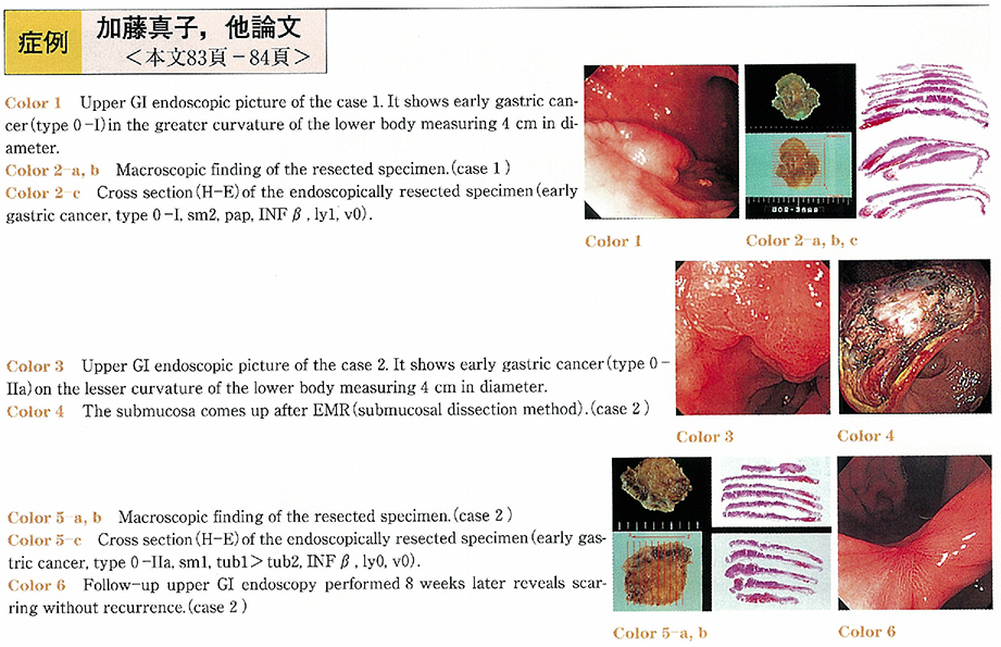
We experienced two early gastric cancers, with over 40 mm in size, resected by a single piece using submucosal dissection EMR.
The first patient was a 61-year-old man with an early gastric cancer (type 0-I) at the greater curvature of the lower body measuring 4 cm in diameter. Biopsy taken from the tumor revealed a papillary adenocarcinoma and endoscopic ultrasonograhy (EUS) demonstrated that the tumor did not invade the submucosa. Submucosal dissection EMR using an electro-surgical snare and a flex knife was performed. The lesion was completely resected with one piece. The size of the resected specimen was 46×45mm. Histological examination showed an early gastric cancer type 0-I (sm2, pap, INFβ, ly1, v0) . Additional surgical resection was carried out, but no metastatic lesion was detected in the regional lymph nodes.
The second patient was an 88-year-old woman with an early gastric cancer (type 0-IIa) at the lesser curvature of the lower body, EUS demonstrated that the tumor did not invade the submucosa. Submucosal dissection EMR was performed in the same way as the first case. The resected specimen was 40×35mm and histological findings showed an early gastric cancer type 0-IIa (sm1, tub1>tub2, INFβ, ly0,v0) . The tumor minimally invaded the submucosa (sm1) with tumor free margins of the resected specimen. Additional surgery was not performed considering her age.
Submucosal dissection EMR makes it possible to remove tumors with one piece regardless of size, which gave us very important pathological findings to determine a complete resection or not.

Fullsize Image
著者関連情報
© 2003 一般社団法人 日本消化器内視鏡学会 関東支部
前の記事 次の記事
feedback
Top